The trouble with uric acid crystals is you know they are there. But you cannot see them.
Also known as gout crystals, these deposits of solid uric acid are too small to see, but devastating in their effects. It’s worrying to know there are millions of tiny particles floating around your body, that remain hidden.
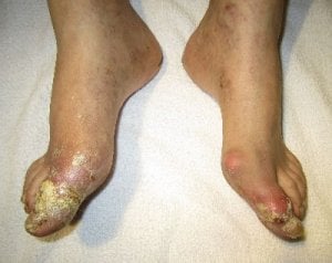
Now, strictly speaking, uric acid crystals don’t always remain hidden. As large deposits form tophi, that can break through the skin. But for our peace of mind, it is a good idea to see what is happening in our joints. Especially as imaging can help our doctors with gout diagnosis and uric acid treatment.
Traditionally, there was only one way to test for the presence of uric acid crystals. That is to analyze fluid from the joints. Recently, there is hope that DECT might eventually replace this invasive procedure, but the huge expense of suitable scanning equipment puts this beyond the reach of most gout patients.
Advances in other medical imaging techniques lead to frequent reviews in professional journals. The most recent[1] of these concludes:
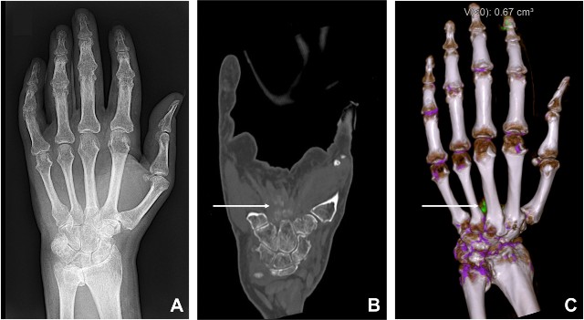
The diagnosis of gout is usually based on clinical presentation and laboratory findings. Imaging plays a role in the assessment and grading of articular damage related to chronic, long-standing disease, which is characterized by granulomatous synovitis, tophi, and erosions. Multimodality imaging of chronic tophaceous gout may be useful in clinical practice for a variety of purposes, including assessment of disease-related anatomical changes and monitoring of articular and soft-tissue lesions over time, especially in response to urate-lowering therapy. Radiography remains the primary imaging technique. Ultrasonography may detect monosodium urate crystals on cartilage, is helpful to assess small joint effusion, to guide to joint aspiration, and to evaluate the volume of tophi. Computed tomography is considered to be more sensitive than plain radiography in the detection and evaluation of cortical bone erosions associated with tophi. MRI represents the only imaging modality which provides visualization of bone marrow edema associated with erosions and may be useful to characterize and distinguish tophi from other soft tissue nodules.
Unfortunately, the full report is in Italian, and impossible for me to understand. However, an earlier review[2] shows similar findings, and so we can see how different technologies play their part in allowing us to see uric acid crystals (or not).

X-ray
Uric acid crystals will not show on x-rays. However, they do show joint damage where gout has eaten into the bone. Unfortunately, this is too late to prevent that damage.
Ultrasound
The relatively low cost, and high availability, of ultrasound, make it the most reliable tool for measuring gout progression. It will detect deposits at quite an early stage, though it will not determine if these are uric acid crystals from gout or other deposits such as pseudogout. If there is any doubt, the ultrasound can also help in positioning the rheumatologist’s needle for joint fluid analysis.
CT Scans
Computed Tomography will detect uric acid crystals at very low volumes. It also shows clearly when these deposits start growing into bone – a much earlier recognition of bone damage compared to x-ray. Though this early detection of deposits allows urate treatment to start before gout causes permanent damage, the scarcity of suitable equipment limits this to the lucky few.
MRI
MRI, though not cheap, is more widely available. It will detect bone erosion earlier than x-ray, and it will detect deposits, though not what they are. The 2009 study concludes that MRI might be most useful in assessing joint problems associated with gout, whilst the later report notes that it is the only technique to show bone marrow swelling [edema].
Leave Uric Acid Crystals to read the Understanding Uric Acid Section.
Uric Acid Crystals Related Topics
Please remember: to find more related pages that are relevant to you, use the search box near the top of every page.
Common Terms: crystal
Other posts that include these terms:
- Gout Without Hyperuricemia
- Uric Acid Crystal Pictures
- Urate Crystals
- Treatment For Gout
- Test For Uric Acid Crystals
- Is Gout Massage A Bad Treatment Of Gout?
- Uric Acid Crystal Formation and Growth
Uric Acid Crystals Imaging References
- Paparo, F., L. M. Sconfienza, A. Muda, A. Denegri, R. Piccazzo, E. Aleo, and M. A. Cimmino. “Multimodality imaging of chronic tophaceous gout.” Reumatismo (2010): 286-291.
- Perez-Ruiz, Fernando, Nicola Dalbeth, Aranzazu Urresola, Eugenio de Miguel, and Naomi Schlesinger. “Gout. Imaging of gout: findings and utility.” Arthritis research & therapy 11, no. 3 (2009): 232.
Please give your feedback
Did this page help you? If yes, please consider a small donation. Your donations help keep GoutPal's gout support services free for everyone.
If not, please tell me how I can improve it to help you more.
- YouTube
- The gout forums.


