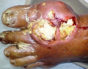
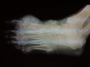
These images are from a summary of a new case study about tophi in the left foot.
This ties in with a recent review of unusual tophi presentation (search for occupational gout)
The working title of my summary is How Bad Can Feet Tophi Get?
I think you’ll understand the title better once you see the images, but first the abstract.
Feet Tophi Case Report: Abstract
- Title:
- Multiarticular chronic tophaceous gout with severe and multiple ulcerations: a case report.
- Authors:
- Falidas E, Rallis E, Bournia VK, Mathioulakis S, Pavlakis E, Villias C.
- Published:
- J Med Case Rep. 2011 Aug 19;5:397.
In my summary, I will use laymans terms to explain the medical terms and issues. Multiarticular means affecting many joints.
Feet Tophi Case Report: Introduction
Gout is a common inflammatory arthritis caused by articular precipitation of monosodium urate crystals. It usually affects the first metatarsophalangeal joint of the foot and less commonly other joints, such as wrists, elbows, knees and ankles.
The first metatarsophalangeal joint is the big toe, or bunion joint. I focus on feet here, but will include other joints in my published summary.
Feet Tophi Case Report: Case Presentation
We report the case of a 75-year-old Caucasian man with tophaceous multiarticular gout, soft-tissue involvement and ulcerated tophi on the first metatarsophalangeal joint of the left foot, on the first interphalangeal joint of the right foot and on the left thumb.
Feet Tophi Case Report: Conclusion
Ulcers due to tophaceous gout are currently uncommon considering the positive effect of pharmaceutical treatment in controlling hyperuricemia. Surgical treatment is seldom required for gout and is usually reserved for cases of recurrent attacks with deformities, severe pain, infection and joint destruction.
Feet Tophi Case Report: Pictures
Feet Tophi Pictures: On Admission

Feet Tophi Pictures: Xray

Feet Tophi Pictures: Resolved
 Complete healing of the ulcer 40 days after the initial observation.
Feet Tophi Case Report: Next Steps
Pretty nasty eh? Can you show worse? You can upload images in the gout forum. You can also discuss this gout research, or any other Gout Study Information.
To stay informed when more feet tophi studies, plus other relevant gout information, are published, please subscribe to the free update service:
Subscribe by clicking this button
Subscription is free and your email address is safe – I will never share it with anyone else.